

Yes, you can receive dental implants even with thin jawbones. Modern surgical techniques allow dentists to rebuild bone volume before or during implant placement. This guide explains how dental implants and oral surgery at EDHAK Center in Banha, Egypt, address atrophic ridges. We cover bone grafting, sinus lifts, specialized implant designs, and the imaging required for safe treatment planning.
Bone Grafting Techniques
Bone grafting is a surgical procedure that adds volume to the jawbone to support dental implants. When natural bone resorbs after tooth loss, the ridge becomes too narrow for standard implants. Grafting restores the necessary width and height. At EDHAK Center, we utilize various grafting materials tailored to the patient's specific bone deficiency.
Autogenous Grafts
Autogenous grafts use bone harvested from the patient's own body, such as the chin or hip. This material is the gold standard because it contains living cells that promote new bone growth. However, it requires a second surgical site, which can increase recovery time. according to Basal Implants An For additional details, review the Dental Services EDHAK Center.
Allografts and Xenografts
Allografts are processed human bone, while xenografts are animal-derived bone, typically bovine. These materials act as scaffolds for new bone formation. They are widely used because they are readily available and do not require a second surgical site.
Alloplasts
Alloplasts are synthetic materials, such as hydroxyapatite or beta-tricalcium phosphate. These materials are biocompatible and predictable. They are often used in combination with other graft types to enhance stability.
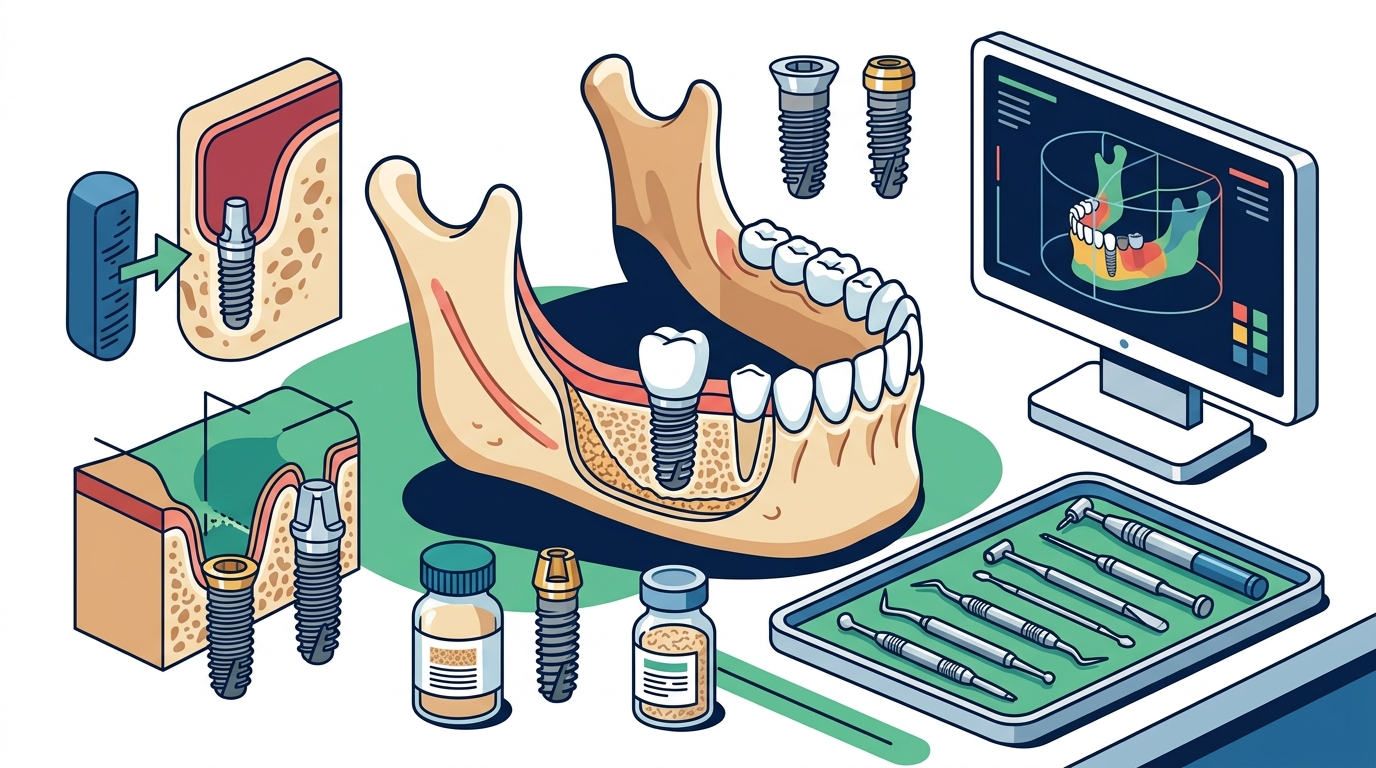
Sinus Lift Procedures
A sinus lift is a procedure that adds bone to the upper jaw, specifically in the molar region. The maxillary sinus sits directly above the upper molars, limiting vertical bone height. When this space is insufficient, a sinus lift elevates the sinus membrane to create room for an implant.
Lateral Window Approach
The lateral window approach involves creating a small opening in the bone wall next to the sinus. The surgeon lifts the sinus membrane and packs graft material underneath. This technique is ideal for patients with significant vertical bone deficiency.
Transalveolar Approach
The transalveolar, or crestal, approach is less invasive. The surgeon drills into the bone crest and gently lifts the sinus membrane from below. This method is suitable for patients with moderate bone deficiency and is often performed at the same time as implant placement.
Clinical Assessment Requirements
Clinical assessment is the first step in determining implant candidacy. It involves a thorough examination of the oral cavity, medical history, and radiographic imaging. Dentists must evaluate bone quality, quantity, and the position of vital structures like nerves and sinuses.
Medical History Review
Patients must disclose all medical conditions, including diabetes, osteoporosis, and smoking habits. These factors can affect bone healing and implant success rates. A comprehensive medical review ensures that the treatment plan is safe and realistic.
Oral Examination
The dentist examines the gums, teeth, and bite alignment. Soft tissue health is critical for long-term implant success. Any active infections or periodontal disease must be treated before implant surgery.
Specialized Implant Designs
Specialized implant designs are engineered to work in challenging bone conditions. Standard implants require a certain amount of bone for stability. When bone is thin, specific designs can provide the necessary support.
Narrow-Diameter Implants
Short Implants
Short implants are used when vertical bone height is insufficient. They are often placed in the posterior maxilla to avoid sinus lift procedures. Short implants have shown high success rates in modern studies.
Specialized Implant Types
Basal Implants
Zygomatic Implants
Zygomatic implants are long implants that anchor into the cheekbone. They are used in patients with severe maxillary atrophy. These implants bypass the need for extensive bone grafting in the upper jaw.
D Imaging Assessment
D imaging assessment refers to the use of digital imaging technologies to evaluate bone structure. Traditional 2D X-rays provide limited information about bone volume. Advanced imaging provides a comprehensive view of the jaw.
Cone Beam CT (CBCT)
Cone Beam CT is the gold standard for implant planning. It provides 3D images of the jaw, allowing precise measurement of bone width, height, and density. CBCT scans help dentists avoid vital structures like the inferior alveolar nerve.
Digital Scans
Digital intraoral scans capture the shape of the teeth and gums. These scans are used to create surgical guides and prosthetic models. They enhance the accuracy of implant placement and restoration.
Bone Grafting
Bone grafting is essential for patients with thin jawbones. It restores the bone volume needed for implant stability. The choice of graft material depends on the size of the defect and the patient's healing capacity.
Guided Bone Regeneration
Healing Time
Bone grafting requires a healing period before implant placement. This period typically ranges from 3 to 6 months. During this time, the graft material integrates with the native bone.
Sinus Lift
Sinus lift procedures are critical for upper jaw implant placement. They increase vertical bone height in the posterior maxilla. The procedure is safe and predictable when performed by experienced surgeons.
Simultaneous vs. Staged
Simultaneous sinus lift and implant placement is possible when there is sufficient primary stability. Staged procedures involve placing the graft first, waiting for healing, and then placing the implant. The choice depends on the amount of bone available.
Zygomatic Implants
Zygomatic implants are a solution for severe maxillary atrophy. They are longer than standard implants and anchor into the zygomatic bone. This technique avoids the need for extensive bone grafting.
Indications
Implant Evaluation Imaging
Implant evaluation imaging is a critical component of treatment planning. It ensures that implants are placed in optimal positions. Accurate imaging reduces the risk of complications and improves long-term outcomes.
3D Planning Software
3D planning software allows dentists to simulate implant placement virtually. This software helps in designing surgical guides and prosthetics. It enhances precision and reduces surgical time.
Ridge Augmentation
Ridge augmentation is a procedure that increases the width of the alveolar ridge. It is often performed in conjunction with bone grafting. This technique is essential for patients with horizontal bone deficiency.
Techniques
Comparison of Bone Augmentation Techniques
| Technique | Indication | Healing Time | Invasiveness |
|---|---|---|---|
| Autogenous Graft | Large defects | 4-6 months | High |
| Xenograft | Moderate defects | 3-6 months | Moderate |
| Sinus Lift | Vertical deficiency | 4-8 months | Moderate |
| Zygomatic Implant | Severe atrophy | Immediate loading | High |
Key Takeaways
- Thin jawbones do not disqualify you from dental implants.
- Bone grafting and sinus lifts are standard procedures for atrophic jaws.
- CBCT imaging is essential for accurate implant planning.
- Zygomatic implants are a viable option for severe maxillary atrophy.
- Healing times vary depending on the procedure and graft material used.
- Consultation with a specialist is crucial for personalized treatment planning.
Frequently Asked Questions
Can I get implants if my jawbone is too thin?
Yes, bone grafting and sinus lift procedures can restore sufficient bone volume for implant placement.
How long does bone grafting take to heal?
Healing typically takes 3 to 6 months, depending on the size of the defect and the graft material used.
What is a sinus lift?
A sinus lift is a procedure that adds bone to the upper jaw by elevating the sinus membrane.
Are zygomatic implants safe?
Yes, zygomatic implants are a safe and effective option for patients with severe maxillary atrophy.
Do I need a CT scan before implant surgery?
Yes, a CBCT scan is recommended to evaluate bone volume and plan implant placement accurately.
Can I eat normally after bone grafting?
You will need to follow a soft diet for several weeks to allow the graft site to heal properly.
Conclusion
Thin jawbones are not a barrier to successful dental implant treatment. With advanced techniques like bone grafting, sinus lifts, and specialized implant designs, patients can achieve stable and functional restorations. At EDHAK Center, we provide comprehensive care for patients with atrophic jaws. Our team in Banha, Egypt, is dedicated to helping you restore your smile with confidence. Contact us today to schedule a consultation and discuss your treatment options.